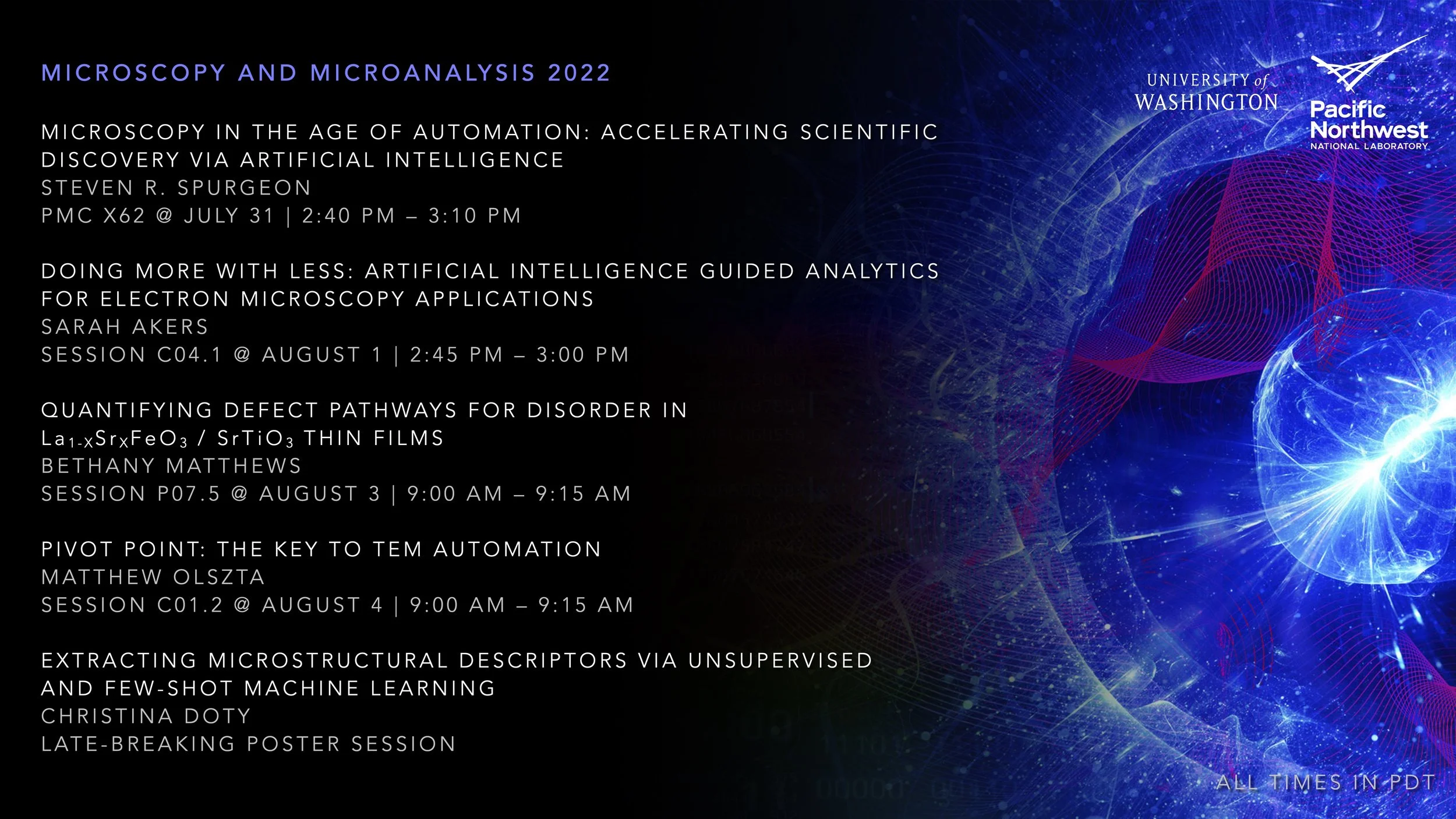
Upcoming M&M 2022 Presentations
Our team is excited to share our research in many talks and posters at the upcoming Microscopy and Microanalysis Meeting in Portland! We will show how first-of-its-kind artificial intelligence can accelerate materials design, automate microstructural analysis, guide high speed in situ decision-making, and reimagine microscope operation. We hope that you will be able to join us!
Microscopy in the Age of Automation: Accelerating Scientific Discovery via Artificial Intelligence
Steven R. Spurgeon
PMC X62 @ July 31 | 2:40 PM – 3:10 PM
Artificial intelligence (AI) promises to reshape scientific inquiry and enable breakthrough discoveries in areas such as energy storage, quantum computing, and biomedicine. While many domains have embraced AI, specifically in the form of automation and machine learning (ML), its adoption for electron microscopy of non-biological systems has been slower. Here I will describe our efforts to develop a framework for data-driven discovery in the electron microscope. I will discuss three topics: (1) emerging sparse data analytics for microstructural classification in real-world scenarios, (2) edge computing-accelerated instrument controllers for self-driving experimentation, and (3) forecasting models for predicting the evolution of in situ data.
Relevant Publication:
Olszta, M., Hopkins, D., Fiedler, K.R., Oostrom, M., Akers, S., and S.R. Spurgeon. “An automated scanning transmission electron microscope guided by sparse data analytics.” Microscopy and Microanalysis. (2022). DOI:10.1017/S1431927622012065 [Download PDF]
Doing More with Less: Artificial Intelligence Guided Analytics for Electron Microscopy Applications
Sarah Akers
Session C04.1 @ August 1 | 2:45 PM – 3:00 PM
Scanning transmission electron microscopy (STEM) is an important tool in the study of microstructures and ultimately a wide range of material and chemical systems. Quantitatively describing these microstructures with advanced artificial intelligence (AI) techniques has recently become an emerging and impactful area of research in the materials domain. Sophisticated new models and algorithms are being developed to automate the task of image characterization, bypassing the need for expensive manual analysis and enabling the discovery of new and unexpected features within an image. We explore the topic of few-shot or low-shot learning (FSL) techniques in the context of creating adaptable AI designed for flexibility in both analytics and data acquisition. FSL, as the name suggests, uses little to no data in training and nearly eliminates the laborious task of data annotation.
Relevant Publication:
Akers, S., Kautz, E., Trevino-Gavito, A., Olszta, M., Matthews, B., Wang, L., Du, Y., and S.R. Spurgeon. “Rapid and flexible segmentation of electron microscopy data using few-shot machine learning.“ npj Computational Materials. 7 (2021): 187. DOI:10.1038/s41524-021-00652-z [Download PDF]
Quantifying Defect Pathways for Disorder in La1-xSrxFeO3 / SrTiO3 Thin Films
Bethany Matthews
Session P07.5 @ August 3 | 9:00 AM – 9:15 AM
Determining the behavior of functional materials under irradiation is important for fundamental understanding of order-disorder phenomena as well as control of performance in extreme conditions for applications such as nuclear reactors or spacecraft. Prior studies of model oxide interfaces have found that certain interface configurations may be more robust to amorphization under irradiation. These findings raise the question of how initial defect distributions with a thin film may affect the evolution of disorder and how such populations can be tuned to guide the radiation response. Here we study the progression of disorder in oxide thin films using analytical scanning transmission electron microscopy, revealing how intrinsic defect populations guide disordering pathways.
Relevant Publication:
Matthews, B., Sassi, M., Barr, C., Ophus, C., Kaspar, T., Jiang, W., Hattar, K., and S.R. Spurgeon. “Percolation of ion-irradiation-induced disorder in complex oxide interfaces.” Nano Letters. 21.12 (2021): 5353–5359. DOI:10.1021/acs.nanolett.1c01651 [Download PDF]
Pivot Point: The Key to TEM Automation
Matthew Olszta
Session C01.2 @ August 4 | 9:00 AM – 9:15 AM
Since its inception, transmission electron microscopy (TEM) has been considered one of the most site-specific sample analysis techniques; with regions of interest on the order to nanometers to micrometers. The advent of aberration Cs probe correction has further pushed the resolution of the scanning TEM (STEM) to the picometer scale, thereby minimizing the desired field of investigation further. Combined with the addition of other factors such time-consuming sample preparation, this trend has minimized the utilization of statistical analysis in extremely high-resolution scenarios. With current automated control of the microscope stage, it is possible to investigate how the stage responds to requests to move to a target location. We will discuss protocols to understand existing stage limitations with respect to stage motion. In order to elicit discussion on both the practicality of current stage designs on full automation as well how next generation microscopes designs to better accommodate advanced microscopy algorithms it is necessary to bring forth these under-appreciated microscopy topics and discuss them in detail.
Relevant Publication:
Olszta, M. and K. Fiedler. “Nanocartography: Planning for success in analytical electron microscopy.” (2022). Arxiv Preprint: https://arxiv.org/abs/2205.03956
Extracting Microstructural Descriptors Via Unsupervised and Few-Shot Machine Learning
Christina Doty
Late-Breaking Poster Session
Scanning transmission electron microscopy (STEM) is a cornerstone of the characterization of microstructures, but features in STEM data are often diverse, artifacted, and poorly described by past libraries. The application of emerging machine learning (ML) techniques to the analysis of STEM images can potentially help researchers discover new features and structures without needing to rely on time-intensive manual analysis. However, many ML models are highly specialized and must be trained on large sets of hand-labeled image data. Generalizing these models and allowing them to adapt to different material systems and conditions without extensive labeled data can be challenging. Here we describe a graphical user interface (GUI)-based application designed to guide microscopists through the training and application of our few-shot ML model. We explore the use of various methods of training-set selection including manual, automated, and a newly developed hybrid unsupervised-few-shot approach.
Relevant Publication:
Doty, C., Gallagher, S., Cui, W., Chen, W., Bhushan, S., Oostrom, M., Akers, S., and S.R. Spurgeon. “Design of a graphical user interface for few-shot machine learning-based classification of electron microscopy data.” Computational Materials Science. 203.15 (2022): 111121. DOI:10.1016/j.commatsci.2021.111121 [Download PDF]
